What is Vet BLUE®?
FAST is an acronym that stands for Focused Assessment with Sonography for Trauma. It is an ultrasound exam developed by trauma surgeons (yes trauma surgeons) in the 1990s and used in people as a screening test for the detection of free fluid in the abdominal cavity, ascites, and the pleural cavity including pleural effusion and pericardial effusion. In 2004 the translational study from humans to dogs was published by Boysen and colleagues out of Tufts.
The next year (2005) we developed both AFAST® and TFAST® as part of my clinical research requirement for my emergency and critical care residency training out of the Emergency Pet Center, San Antonio, Texas. TFAST® was the first FAST protocol developed for the thorax in veterinary medicine. In fact, our TFAST® protocol exceeded the protocol for people called EFAST (“E” for “extended”). Through the detection of lung pathology during TFAST®, we extended additional views beyond its Chest Tube Site view for a more comprehensive lung screening test in 2010, over a decade ago. We named the protocol Vet BLUE®, BLUE an acronym for Brief Lung Ultrasound Exam or Brief Lung Ultrasound in Emergency. We were the first in the world to use lung ultrasound as a proactive part of the physical exam.
Vet BLUE® is a unique regional, pattern-based approach that has its views named caudodorsal, perihilar, middle, and cranial lung regions plus the Diaphragmatico-Hepatic (DH) view. This approach is analogous to how we are taught to interpret lung on radiography. Moreover, we developed a B-line scoring system that inherently provides a Vet BLUE® severity score by combining B-line scoring and their distribution. For signs of consolidation, we have defined several signs including the Shred Sign, Tissue Sign, Wedge Sign, and Nodule Sign. The former 2 signs are borrowed from Dr. Daniel Lichtenstein’s approach in human medicine; however, the Wedge Sign and Nodule Sign are our developed published terms in our peer-reviewed literature. The fundamental orientation for all lung ultrasound is the Gator Sign representing rib-intercostal space-rib.
Global FAST® is the combination of AFAST®, TFAST®, and Vet BLUE® as a single ultrasound format that serves as an extension of the physical exam. We advocate for the use of Global FAST® for all patient point-of-care ultrasound assessment because such an approach prevents “satisfaction of search error” and “confirmation bias error” through selective or targeted-of-care ultrasound imaging, i.e., focused cardiac ultrasound, focused gallbladder, focused urinary tract.

Figure. Fundamental “Gator Sign” Orientation for all Lung Ultrasound. This material is reproduced with permission of John Wiley & Sons, Inc., Point-of-Care Ultrasound Techniques for the Small Animal Practitioner, 2nd Edition, Wiley ©2021 and Greg Lisciandro, Hill Country Veterinary Specialists, FASTVet.com.
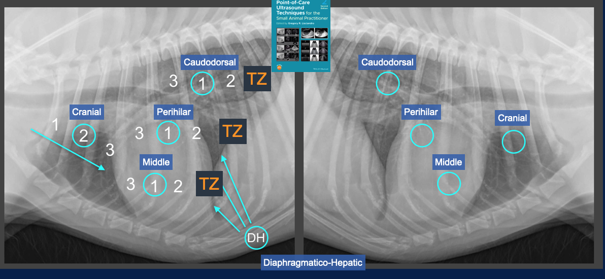
Figure. Vet BLUE® and Its 9-acoustic Windows. Vet BLUE® has the following regional views – caudodorsal, perihilar, middle and cranial lung regions. Each regional view has a minimum of 3 intercostal spaces evaluated. The Diaphragmatico-Hepatic view is also used as a deep window for lung inaccessible from transthoracic views. This material is reproduced with permission of John Wiley & Sons, Inc., Point-of-Care Ultrasound Techniques for the Small Animal Practitioner, 2nd Edition, Wiley ©2021 and Greg Lisciandro, Hill Country Veterinary Specialists, FASTVet.com.
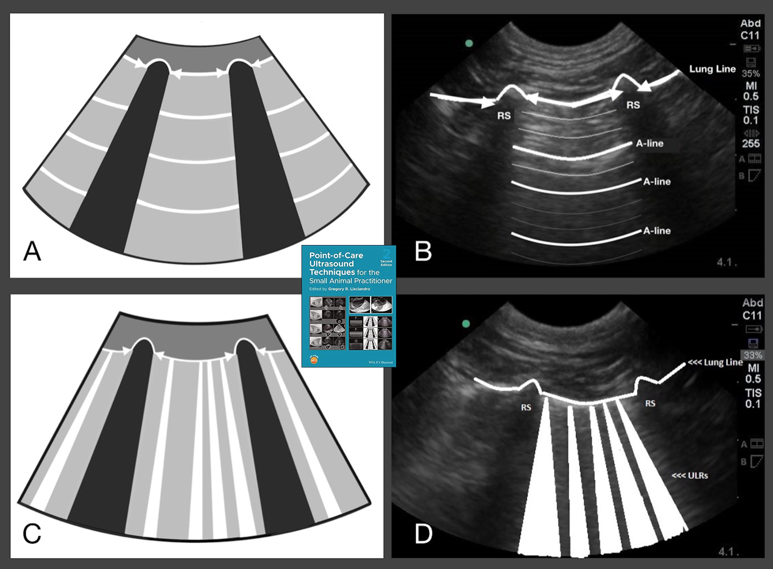
Figure. Dry Lung and Wet Lung. This material is reproduced with permission of John Wiley & Sons, Inc., Point-of-Care Ultrasound Techniques for the Small Animal Practitioner, 2nd Edition, Wiley ©2021 and Greg Lisciandro, Hill Country Veterinary Specialists, FASTVet.com.

Figure. B-line Scoring System. We expect dry lung all views with uncommon single B-lines in dogs and cats and likely all adult mammalian lung. The B-line scoring is 1, 2, 3 as “weak positives” and >3, and infinity, “strong positives” taken as the highest number over a single intercostal space at each respective Vet BLUE® regional view. B-lines and their distribution are a Vet BLUE® inherent severity scoring system and may also be used as a guide to loop diuretic therapy in left-sided congestive heart failure and as a tracking tool. This material is reproduced with permission of John Wiley & Sons, Inc., Point-of-Care Ultrasound Techniques for the Small Animal Practitioner, 2nd Edition, Wiley ©2021 and Greg Lisciandro, Hill Country Veterinary Specialists, FASTVet.com.
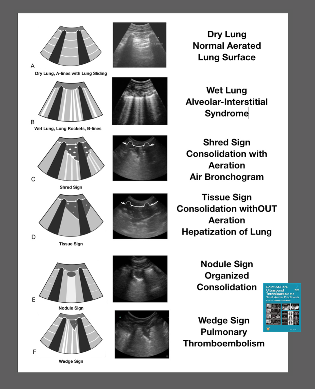
Figure. Vet BLUE® Visual Lung Language and Its 6 Signs. Vet BLUE® Visual Lung Language from most to least normal, from less severe to more severely affected lung, is as follows: Dry Lung to Wet Lung (B-lines, alveolar-interstitial edema) to Shred Sign (air bronchogram) to Tissue Sign (hepatization of lung) to Nodule Sign to Wedge Sign (pulmonary thromboembolism). This material is reproduced with permission of John Wiley & Sons, Inc., Point-of-Care Ultrasound Techniques for the Small Animal Practitioner, 2nd Edition, Wiley ©2021 and Greg Lisciandro, Hill Country Veterinary Specialists, FASTVet.com.
When Should You Perform Vet BLUE®?
The mindset is to use Vet BLUE® as “an extension of the physical exam.” In other words, it’s everyday ultrasound for nearly every patient. A Vet BLUE® with proper training and experience is a < 2-minute screening imaging test that is achievable for the non-imaging specialist with minimal ultrasound training.
Indications for a Vet BLUE®
- Any abnormal or questionable patient
- Your new preanesthetic screening test
- Perioperative monitoring
- Semiannual and annual visits
- Geriatric screening test
- During patient rounds and recheck exams
- Tracking clinical course of pneumonias (Bacterial, fungal, viral, verminous)
- Tracking clinical course of congestive heart failure
- Ruling out all wet lung conditions including cardiogenic and non-cardiogenic edema
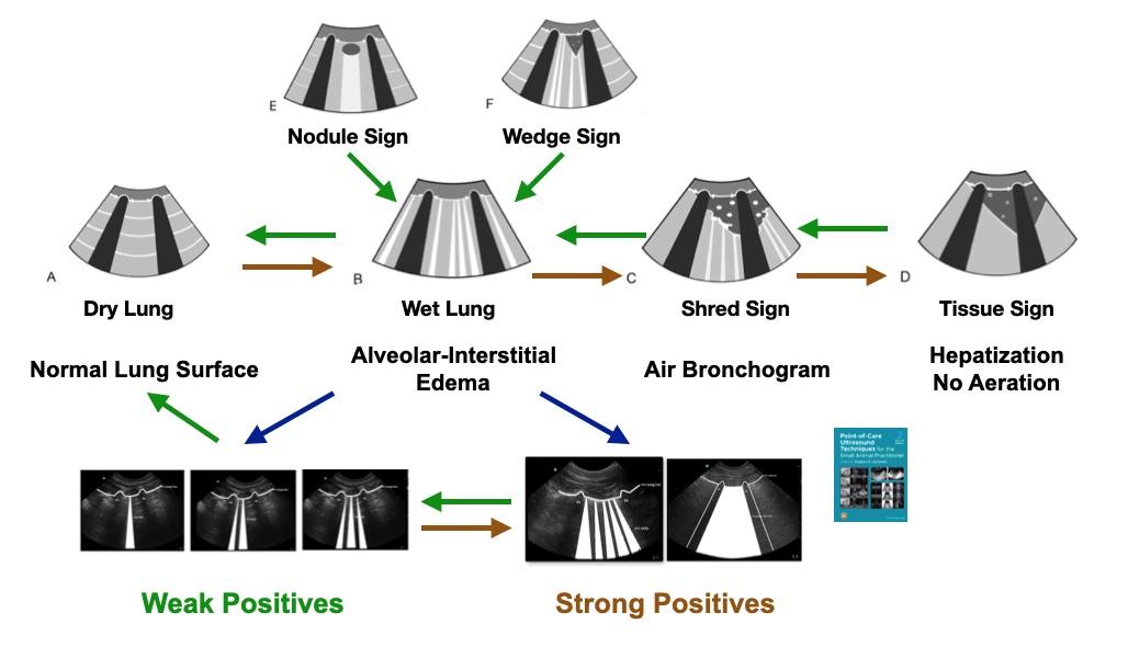
Figure. Use of Vet BLUE® for Tracking Respiratory Conditions. Pneumonia would fall into the 2-way horizontal track of Dry to Wet to Shred Sign to Tissue Sign. B-lines may further be subdivided into “weak” and “strong” positives. This material is reproduced with permission of John Wiley & Sons, Inc., Point-of-Care Ultrasound Techniques for the Small Animal Practitioner, 2nd Edition, Wiley ©2021 and Greg Lisciandro, Hill Country Veterinary Specialists, FASTVet.com.
Advantages of Vet BLUE®
- TFAST® is rapid, radiation sparing, low impact (minimal restraint), and real-time information (no delay) right there at your patient’s side especially when transport and patient status make radiography too risky. With proper training and minimal experience and Vet BLUE® exam should take < 2-minutes.
- Vet BLUE® is more sensitive that thoracic radiography (TXR) for wet lung conditions and performs close to the gold standard test of computed tomography (CT).
Vet BLUE® is extremely sensitive for detecting lung surface pathology and helps better interpret TXR and CT findings.
Vet BLUE®: Recording Findings
Goal-directed templates (GDTs) for recording your findings are a must for success. They keep you disciplined and on task. Vet BLUE® is performed the same order every time just like a cardiologist and a radiologist perform their ultrasound examinations the same way every time. GDTs also give you value for comparison to future exams for you and for your colleagues and show your Vet BLUE® objectives.
Examples of GDTs may be found at our website FASTVet.com on our Free Resources page and by clicking here.
References
- Lisciandro GR, Puchot ML, Gambino JM, Lisciandro SC. The Wedge Sign: A Possible Lung Ultrasound Sign for Pulmonary Thromboembolism. J Vet Emerg Clin Care 2022;32(5):663-669.
- Pacholec C, Lisciandro GR, Masseau I, et al. Lung ultrasound nodule sign for detection of pulmonary nodule lesions in dogs: Comparison to thoracic radiography using computed tomography as the criterion standard. Vet J 2021 Jul 31;105727. doi: 10.1016/j.tvjl.2021.105727. Online ahead of print.
- Ward JL, Murphy SD, Lisciandro GR, et al. Comparison of curvilinear-array (microconvex) and phased-array transducers for ultrasonography of the lungs in dogs. Am J Vet Res, In Press, 2021.
- Dicker SA, Lisciandro GR, Newell SM, et al. Diagnosis of pulmonary contusions with point-of-care lung ultrasonography and thoracic radiography compared to thoracic computed tomography in dogs with motor vehicle trauma: 29 cases (2017-2018). J Vet Emerg Crit Care, 2020 30(6):638-646.
- Vientós-Plotts AI, Wiggen KE, Lisciandro GR, et al. The utility of point-of-care ultrasound non-echo right-sided cardiac markers as a screening test for moderate to severe pulmonary hypertension in dogs. Vet J 2019; 250:6-13.
- Ward JL, Lisciandro GR, Ware WA, Miles KG, DeFrancesco TC. Lung ultrasound findings in 100 dogs with various etiologies of cough. J Am Vet Med Assoc 2019:255(5):574-583.
- Ward JL, Lisciandro GR, Ware WA, et al. Evaluation of Point-of-care Thoracic Ultrasound and NT-proBNP for the Diagnosis of Congestive Heart Failure in Cats with Respiratory Distress. J Vet Intern Med 2018; 32(5): 1530-1540.
- Ward JL, Lisciandro GR, DeFrancesco TD. Distribution of alveolar-interstitial syndrome in dogs and cats with respiratory distress assessed with lung ultrasound versus thoracic radiographs. J Vet Emerg and Crit Care 2018; 28(5): 415-428.
- Lisciandro GR, Fulton RM, Fosgate GT, Mann KA. Frequency and number of B-lines using a regionally-based lung ultrasound examination in cats with radiographically normal lung compared to cats with left-sided congestive heart failure. J Vet Emerg Crit Care 2017; 27(3):267-277.
- Ward JL, Lisciandro GR, Tou SP, Keene BW, DeFrancesco TC. Accuracy of point-of-care lung ultrasound (Vet BLUE protocol) for the diagnosis of cardiogenic pulmonary edema in dogs and cats with acute dyspnea. J Am Vet Assoc 2017; 250(6):666-675.
- Lisciandro GR, Fosgate GT, Fulton RM. Frequency of ultrasound lung rockets using a regionally-based lung ultrasound examination named veterinary bedside lung ultrasound exam (Vet BLUE) in 98 dogs with normal thoracic radiographic lung findings. Vet Rad Ultrasound 55(3):315-22.
- Lisciandro GR. Focused abdominal (AFAST) and thoracic (TFAST) focused assessment with sonography for trauma, triage and monitoring in small animals. J Vet Emerg Crit Care 2011;20(2):104-122.
- Lisciandro GR. Chapter 22: POCUS: Vet BLUE-Introduction and Image Acquisition. In Point-of-care Ultrasound Techniques for the Small Animal Practitioner, 2nd Edition, Ed. Lisciandro GR. Wiley Blackwell: Ames IA 2021.
- Lisciandro GR. Chapter 23: POCUS: Vet BLUE-Clinical Integration, Point-of-care Ultrasound Techniques for the Small Animal Practitioner, 2nd Edition, Wiley-Blackwell: St. Louis, ©2021.
- Lisciandro GR. Cageside Ultrasonography in the Emergency Room and Intensive Care Unit. Vet Clin North Am Small Anim Pract 2020;50(6):1445-1467.
- Lisciandro GR. Chapter 3: Point-of-Care Ultrasound. In Small Animal Diagnostic Ultrasound, 4th Edition, Eds. Mattoon JS, Sellon R, Berry CR. Elsevier: St. Louis MO, 2021.
- Chou Y, Ward JL, Baron LZ, Murphy SD, Topf MA, Lisciandro GR, et al. Focused ultrasound of the caudal vena cava in dogs with cavitary effusions or congestive heart failure: a prospective observational study. PLoS One 2021;16(5):e0252544. doi:10.1371/journal.pone.0252544
- Lisciandro GR, Gambino JM, Lisciandro SC. Case series of 13 dogs and 1 cat with ultrasonographically-detected gallbladder wall edema associated with cardiac disease. J Vet Intern Med 2021 35(3):1342-1346.