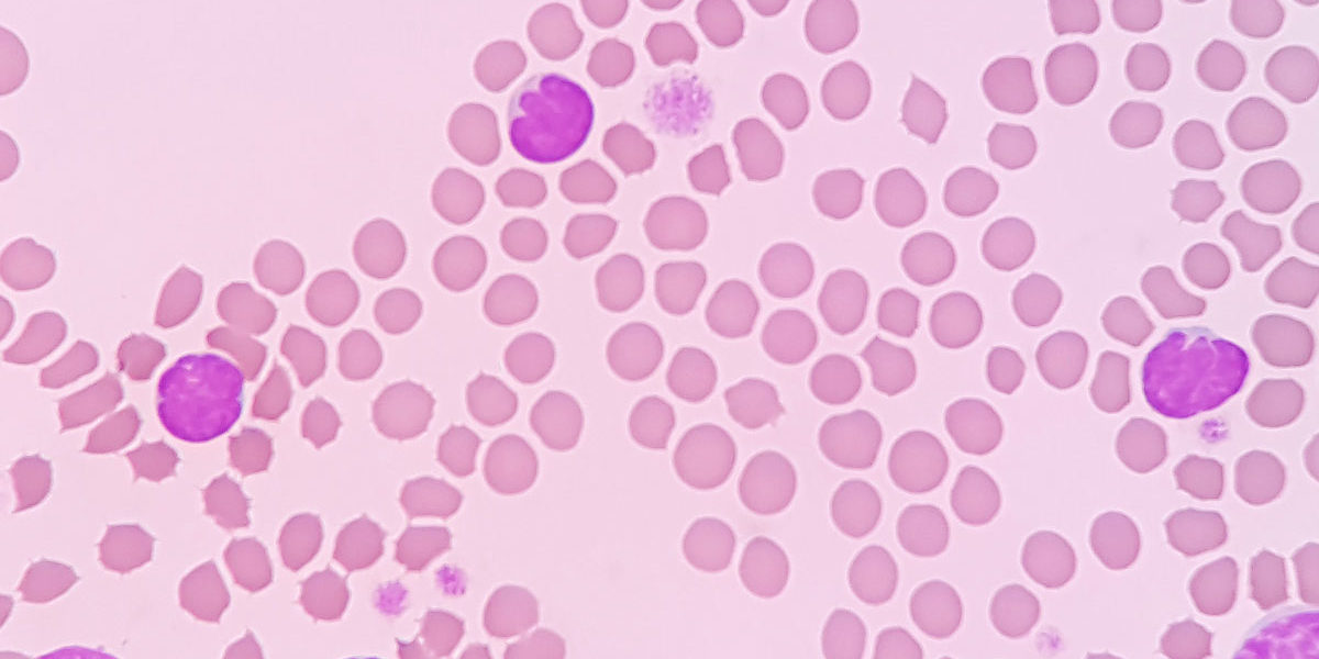
A complete blood count is a simple and fast test routinely performed as a basic part of a health assessment. Evaluation of red blood cell morphology is an important part of a CBC. Changes detected in red blood cells can indicate infections, neoplasia, poisoning, or metabolic disorders and help guide the choice of additional diagnostic tests.
The list below and the accompanying infographic include some of the changes seen in red blood cell morphology and their potential causes.
Normal – Normal erythrocytes are non-nucleated, spherical small cells with a relatively dark rim and central pallor.
Acanthocytes (spur cells) – These irregular, often club-like projections can be found with hepatic lipidosis or hemangiosarcoma.
Echinocytes (crenation, burr cells) – These cells have multiple spine-like projections that can form in response to liver disease or lymphosarcoma. They may also be an artifact of sample preparation.
Drepanocytes (sickle cells) – These crescent-shaped cells can be a normal finding in deer and Angora goats.
Spherocytes – Spherocytes lack central pallor. They may be a consequence of hemolytic anemia or a transfusion mismatch.
Keratocytes (helmet cells, blister cells) – These cells with 2 projections toward what looks like a hole in the cell can be found in cases of hemangiosarcoma or other types of neoplasia, glomerulonephritis, or liver disease.
Schistocytes – Schistocytes are fragmented red blood cells with pointed projections and may be caused by DIC, hemangiosarcoma, or iron deficiency.
Basophilic stippling – This feature (small to large blue inclusions throughout the cell) may be due to residual DNA in the erythrocyte or a response to lead poisoning or anemia.
Howell-Jolly bodies – These small, dark purplish inclusions may represent nuclear remnants or be seen in response to anemia.
Heinz bodies – Heinz bodies are small clumps that often protrude from the cell surface. They may be formed by denatured hemoglobin and can be found in cases of hemolytic anemia, lymphosarcoma, hyperthyroidism, or diabetes mellitus. They can also be a normal feature in the hemogram of cats.
Macrocytes – These large cells are young erythrocytes and are typically found in cases of regenerative anemia. They result from the bone marrow releasing red blood cells into the circulation more quickly than usual.
Nucleated erythrocytes (rubricyte, metarubricyte) – These cells with a large, dark-staining nucleus are also immature cells that have been released into the circulation and are also commonly seen as part of the response to anemia.
Codocytes (target cells) – These cells with a central mass of hemoglobin, a pale middle region, and a dark outer rim are usually associated with liver disease or hypothyroidism.
Other Morphologic Changes in Red Blood Cells
In addition to the changes described above, other red blood cell types with altered morphology include eccentrocytes, hypochromic cells, ovalocytes, polychromatophils, cells with siderotic inclusions, and stomatocytes. Eccentrocytes include dense-staining hemoglobin that is found near a pale zone in the cell. This is a sign of oxidative damage and is often associated with hemolysis. Hypochromic cells have increased central pallor and may indicate iron deficiency. Ovalocytes have an ovoid or elliptical shape. In cats, they are associated with hepatic lipidosis and bone marrow abnormalities. In dogs, they have been linked to myelofibrosis, glomerulonephritis, and myelodysplastic syndrome. Polychromatophils are large, bluish-red cells. They are immature red blood cells and may be seen in low numbers in normal dogs and cats. Increased numbers indicate a response to hypoxia or a regenerative response to anemia. Siderotic inclusions (siderocytes) are small basophilic inclusions that are found near the edge of the cell. They can be associated with transfusions, lead or zinc poisoning, myeloid neoplasia, or some inflammatory disorders. Stomatocytes have an oval or elongated area of central pallor. They are often an artifact, but may also be present when there is a regenerative response to anemia. They can also be found in certain inherited disorders.
Infectious Diseases Targeting Red Blood Cells
Examination of erythrocytes on a blood smear can also reveal infectious agents in dogs, cats, and other domestic animals. Diseases that are caused by red blood cell pathogens include anaplasmosis (affecting domestic and wild ruminants), babesiosis, cytauxzoonosis, mycoplasmosis, and trypanosomosis. Several Babesia spp, may infect dogs and cats, but most species of this genus infect ruminants. They can be seen as intracellular parasites of red blood cells, but the diagnosis is typically confirmed by serology. In cats, Cytauxzoon felis is transmitted by ticks and has been found in midwestern and southern states. Depending on the life stage of the pathogen, it may be found in either white or red blood cells. They can look similar to Mycoplasma spp, Howell-Jolly bodies, or stain artifacts. Depending on the pathogen, Mycoplasma spp may infect dogs, cats, pigs, or several types of ruminants. On a blood smear, they appear as small basophilic rod, round, or ring-shaped structures. Diseases caused by Trypanosoma spp affect all species of domestic animals. Trypanosomes are large, extracellular parasites and are often visible on a blood smear. They can also be found by looking for the motile parasites on an unstained wet mount.
Additional Information
More information on changes in red blood cell morphology and how to prepare a slide and interpret the findings seen on a blood smear can be found at the following sources:

